

Medprime Microscope
Compact & Fully-Automated Bench Top Analyzer
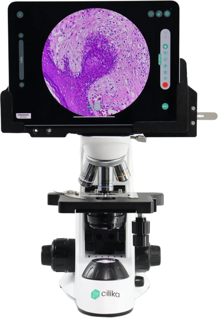
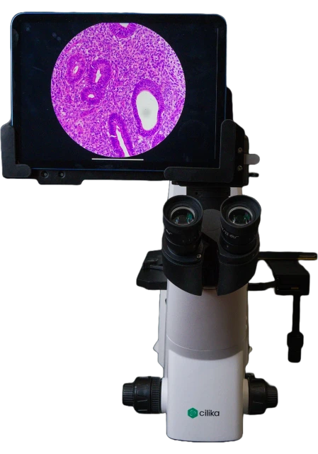
Cilika Digital Microscope
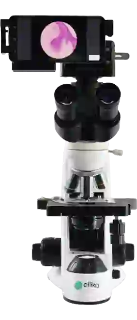
Tablet and Smartphone-Integrated Digital Microscope for Clinical and Research Applications.
Powered by a patented redesigned optical system with TrueView Technology, offering a 100% field of view and zero-lag performance
Backed by over a decade of expertise and trusted by over 1,000 users nationwide
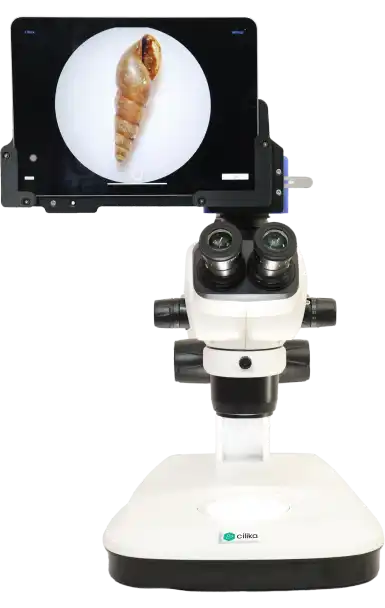
Cilika digital microscope is a combination of tablet/smartphone technology with microscopy. With its redesigned optical system with patented technology, it provides 100% Field of View and zero lag in operations, making it the ideal replacement for traditional microscopes in routine use.
Low Consumption
Only need three reagents. Sample consumed ≤20uL
Efficient
60 samples per hour
Lightweight
390mm(L)×285mm(W)×390mm(H) Weight≤16Kg
Products Highlight
Get to Know CILIKA
Tablet/Smartphone integrated Digital Microscope
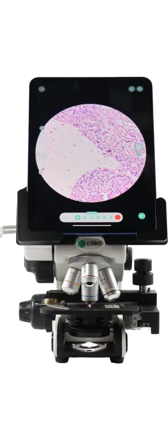
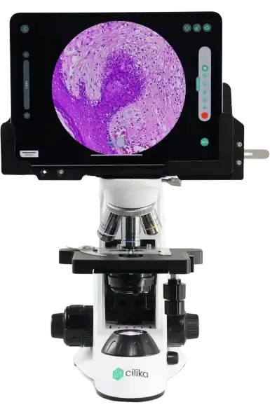
- More than just a digital camera attachment, this is a fully functional iPad integration for seamless operation.
- Featuring a redesigned optical system with patented technology.
100% Field of View
Experts can view the entire circular field of view on the display, just like traditional eyepieces. Unlike conventional camera-based digital microscopes, there is no reduction in the field of view.
Zero lag in Operation
- Smooth transition of the field on display when moving the slide.
- Due to the real-time movement on the display, diagnosing becomes easier and more efficient. As a result, Cilika digital microscope is highly valuable for routine diagnosis.
Ergonomic design to avoid health issues
- Cilika microscopes are designed with precision, taking into account the long-term occupational hazards of microscope use.
- This design prioritizes user comfort, ensuring a safe and efficient experience, making them ideal for extended use without compromising performance.
Inbuilt CILIKA View Software Image analysis
The preloaded Cilika software is specifically designed for Cilika microscopes, enabling micrometry and annotations directly during diagnosis.
- Digital Zoom (up to 5x)
- Calibration and Customization
- Clean User Interface (UI)
- Annotations and Micrometry
- Report Printing with Evidence
High-Quality imaging
- With Cilika, you can capture, edit, and export high-resolution images directly through our app. Adjustable image resolutions of up to 4128×3096 pixels, along with multiple color, contrast, and brightness settings, ensure the perfect image for publications and posters. The white LED illumination provides uniform lighting for optimal clarity.
- Featuring 1500x magnification and digital zoom.
Multiple Device Connectivity and Projection
Cilika microscopes can be easily connected to a projector, allowing multiple users to view samples simultaneously on a digital display. No more waiting for turns everyone sees the same image in real time with consistent clarity. This enhances collaborative learning, group analysis, and telepathology discussions, making microscopy more efficient and interactive.
Compact & Fully-Automated Bench Top Analyzer
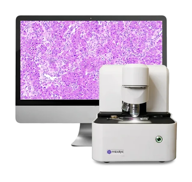
Micalys Whole Slide scanner
Micalys is an automated digital microscope and whole slide scanner, delivering advanced imaging and telepathology solutions for hospitals, laboratories, and educational institutions
Whole slide imaging at 100x objective, Z-stacking capabilities, and enhanced telepathology with Quick Share, Secure IP Sharing, and Live View with Robotic Remote Control
A powerful and reliable whole slide scanner, trusted by professionals to deliver exceptional results
Low Consumption
Only need three reagents. Sample consumed ≤20uL
Efficient
60 samples per hour
Lightweight
390mm(L)×285mm(W)×390mm(H) Weight≤16Kg
Products Highlight
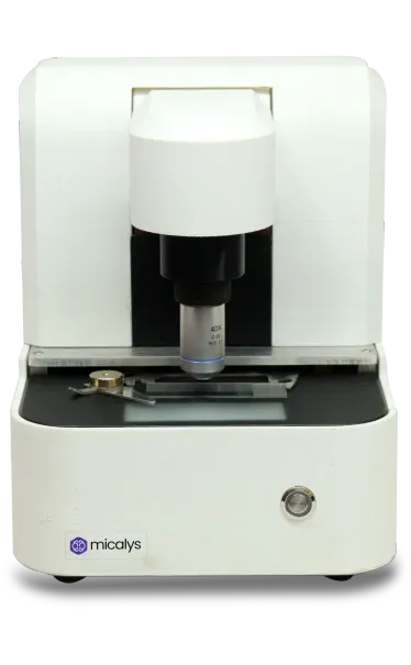
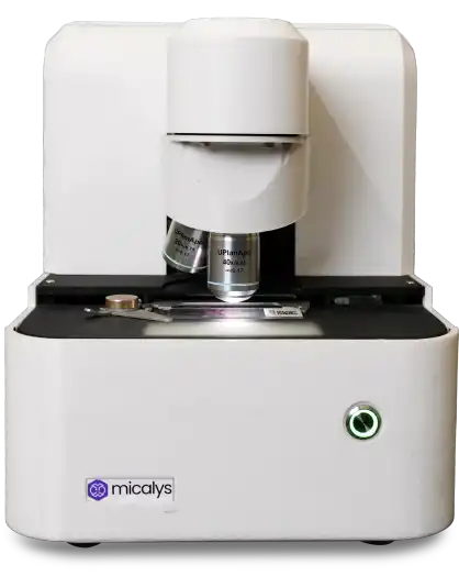
MICALYS S1
Single slide single lens
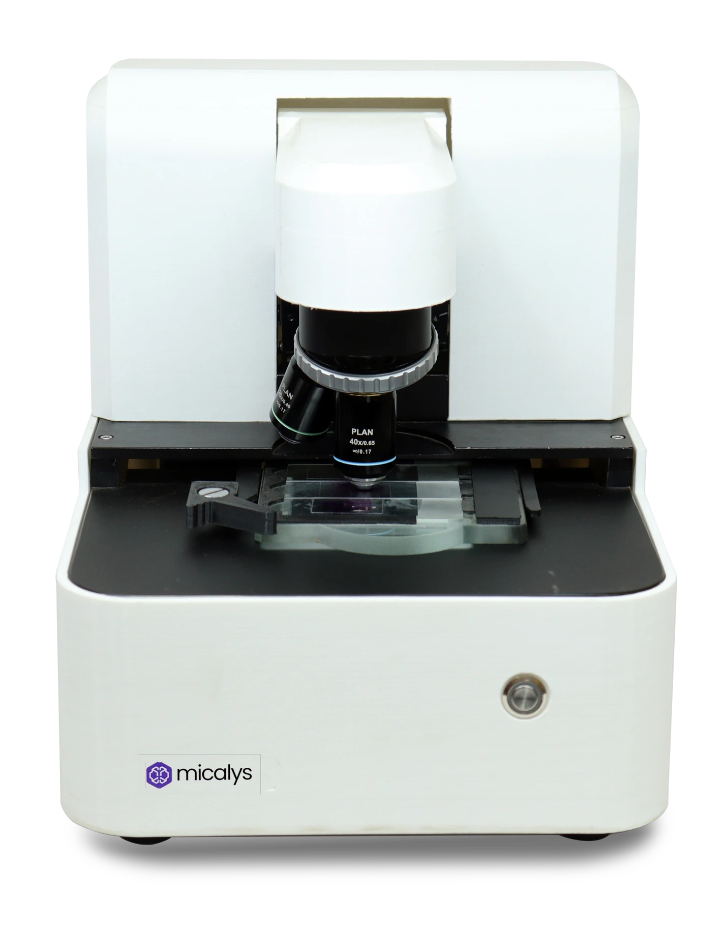
Micalys M1
Single slide multi-lens
Micalys M3
Triple slide multi-lens
Get to Know Micalys
WSI at 100x objective
- Beyond standard 20x and 40x magnification, this scanner features true 100x oil immersion magnification, providing unparalleled clarity and detail.
- Perfect for studying microbiology samples, it allows you to observe every structure closely and effectively.
High resolution Imaging
- High-resolution imaging with fast scanning time enables rapid, precise diagnoses in pathology.
- This technology minimizes misdiagnoses, reduces the need for repeated tests, and supports high throughput for quicker reporting.
11 layers of z stacking
- No need to worry if the slides are uneven, poor quality, or thick. With 11 layers of Z-stacking, the scanner can capture multiple focal planes for clear, detailed imaging.
- This feature ensures you gain insights from every layer, leaving no room for information loss. It’s especially useful for cytology samples.
Boost to telepathology Second opinions made easy
- Quick Share: Generates a link for easy sharing, perfect when you need a second opinion from a colleague or want to quickly share a case for review—no complications, just fast and simple
- Secured IP Sharing: Allows you to securely and easily share findings with your team. Ideal for teams that prefer online diagnosis, ensuring safe and seamless collaboration.
Live view with robotic remote control
- More than just a live view, Micalys lets you remotely manage the microscope’s stage, and focus, and provides magnification with an astonishing latency of just 250 milliseconds.
- This allows you to work anytime, anywhere, without the need to physically carry the microscope or be present in your lab.
Manual Microscopy
- Use the same microscope as a regular digital microscope with the joystick
- Helpful when WSI can’t be performed on non-fixed samples like urine, sperm, or stool, or when the sample is damaged
AI Assistance
- AI-assisted diagnosis for PBS, Malaria, and Filaria provides advanced support in detecting and analyzing samples
- The integration of AI ensures future-ready capabilities, keeping diagnostic processes aligned with the latest advancements.