

Xera X-ray System
Compact & Fully-Automated Bench Top Analyzer
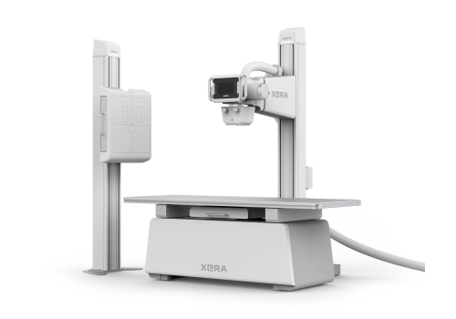
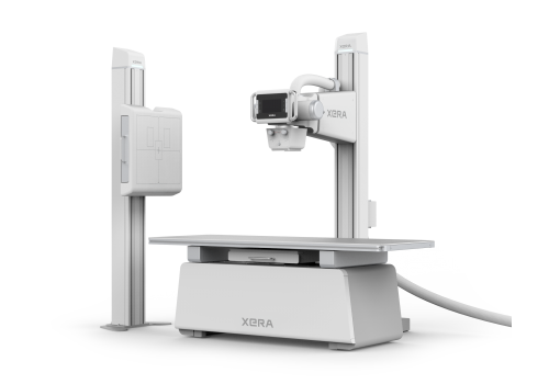
Digital Radiography (X-ray) System
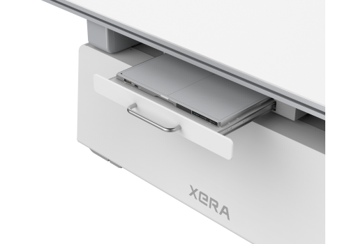
XERA GR10X-40K
Our digital radiography system is aimed to provide a better medical examination experience for humans and animals with its intuitive design and streamlined workflow.
Equipped with cutting-edge technology that achieves high-image quality, improved system usability, and compact design.
The digital radiography system delivers a fast and convenient work experience in your busy hospital environment for an affordable total investment.
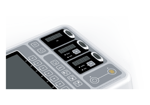
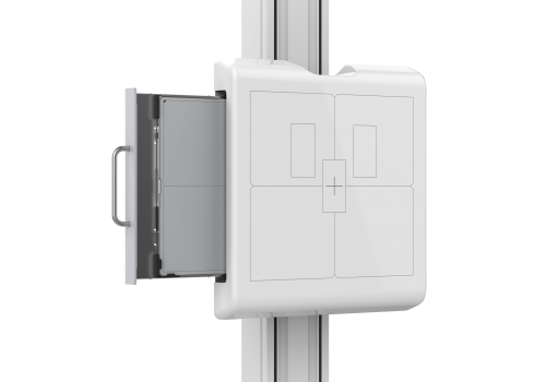
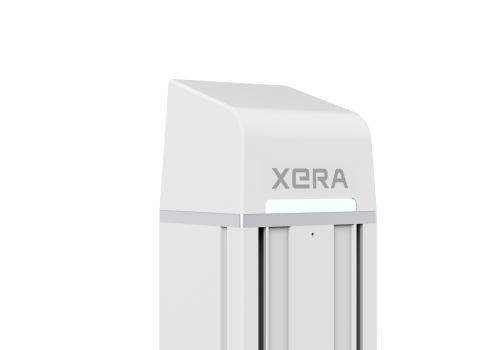
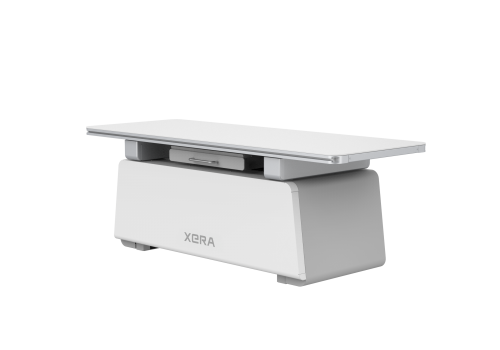
Smart & Ergonomic Design Features
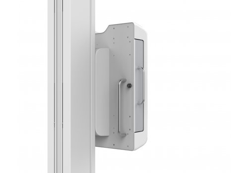
- Height-adjustable chest bucky with smooth tracks for detector change
- LED light at the top of bucky stand as status indicator
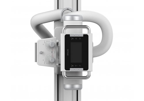
- LCD display monitor
- Wheel controller on console for easy use
- Rotatable stand console handle for better mobility
- Slidable table with curved edges for better safety and mobility
Specifications
Model Name
GR10X-40K
Generator Power Input
230VAC ± 10 %, 50/60 Hz
400 – 480 VAC, 50/60 Hz
Generator Power Output
40kW, 1 phase & 3 phrase
Generator kV Range
40-150 kV
Generator mA range
| 10-500 mA |
Generator mAs range
| 0.1 – 630 mAs |
CE Mark
CE0197
Tube kV Range
40 -125 kV
Tube Rotation
Rotating 16° at high speed
Clinical Performance
Sensitivity
95.58%
Specificity
99.47%
Accuracy
98.01%
Low Consumption
Only need three reagents. Sample consumed ≤20uL
Efficient
60 samples per hour
Lightweight
390mm(L)×285mm(W)×390mm(H) Weight≤16Kg

Contents
Package specifications: 25 T/kit
- SARS-CoV-2 antigen test Cassette
- Extraction tube with sample extraction solution and tip
- Disposable dropper
- IFU: 25 piece/kit
- tubu stand: 1 piece/kit
- Paper cup: 25 piece/kit
- Additional required material: clock/ timer/ stopwatch
- Note: Do not mix or interchange different batches of kits.
- Additional required material: clock/ timer/ stopwatch
- Note: Do not mix or interchange different batches of kits.
Specifications
- Test Item
- Sample Type
- Storage Condition
● SARS-CoV-2 antigen
● Methodology
● Colloidal Gold
● Saliva
● Test Time
● 15mins
● 2-30℃
● Shelf Life
● 24 months
Test Procedure
Preparing
The specimens to be tested and the required reagents shall be removed from the storage condition and be balanced to room temperature; The kit shall be removed from the packaging bag and placed flat on a dry bench.
Testing
- 2.1 Place the test kit horizontally on the table.
- 2.2 Add specimen
Shaking the tube 3 to 5 times and invert the tube so that it is perpendicular to the sample hole (S) and add 3 drops (about 100ul ) of the sample. Set timer for 15 minutes.
- 2.3 Reading the result
The positive specimens can be detected at 15 minutes after sample addition.
Interpretation of Results
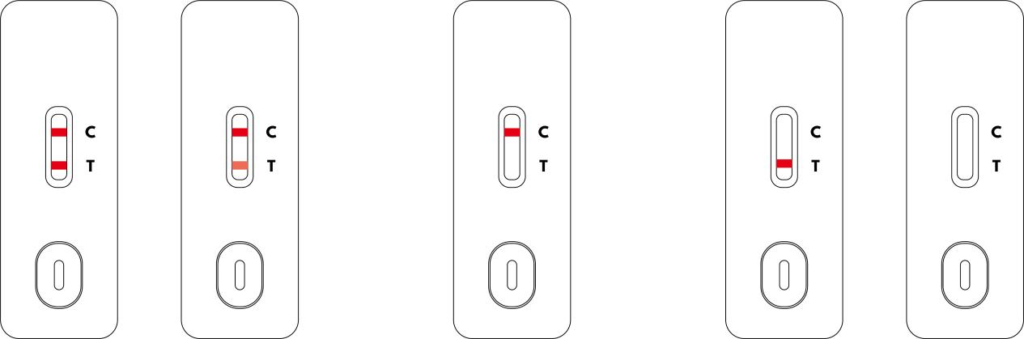
POSITIVE
Two colored lines appear on the membrane. One line appears in the control region (C) and the other line appears in the test region (T).
NEGATIVE
Only one colored line appears in the control region (C). No apparent colored line appears in the test region (T).
INVALID
Control line fails to appear. Results from any test which has not produced a control line at the specified read time must be discarded. Please review the procedure and repeat with a new test. If the problem persists, discontinue using the kit immediately and contact your local distributor.
NOTE
The color intensity in the test region (T) may vary depending on the concentration of analytes present in the specimen. Therefore, any shade of color in the test region should be considered positive. Note that this is a qualitative test only, and cannot determine the concentration of analytes in the specimen. Insufficient specimen volume, incorrect operating procedure or expired tests are the most likely reasons for control line failure.
Compact & Fully-Automated Bench Top Analyzer
Digital Radiography (X-ray) System
XERA GR10X-50K
Our digital radiography system is aimed to provide a better medical examination experience for humans and animals with its intuitive design and streamlined workflow.
Equipped with cutting-edge technology that achieves high-image quality, improved system usability, and compact design.
The digital radiography system delivers a fast and convenient work experience in your busy hospital environment for an affordable total investment.
Smart & Ergonomic Design Features
- Height-adjustable chest bucky with smooth tracks for detector change
- LED light at the top of bucky stand as status indicator
- LCD display monitor
- Wheel controller on console for easy use
- Rotatable stand console handle for better mobility
- Slidable table with curved edges for better safety and mobility
Specifications
Model Name
GR10X-50K
Generator Power Input
400 – 480 VAC, 50/60 Hz
Generator kV Range
40 – 150 kV
Generator mA range
10 – 630 mA
Generator mAs range
0.1 – 630 mAs
CE Mark
CE0197
Tube kV Range
40 – 150 kV
Tube Rotation
Rotating 12° at high speed
Clinical Performance
Sensitivity
95.58%
Specificity
99.47%
Accuracy
98.01%
Low Consumption
Only need three reagents. Sample consumed ≤20uL
Efficient
60 samples per hour
Lightweight
390mm(L)×285mm(W)×390mm(H) Weight≤16Kg
Contents
Package specifications: 25 T/kit
- SARS-CoV-2 antigen test Cassette
- Extraction tube with sample extraction solution and tip
- Disposable dropper
- IFU: 25 piece/kit
- tubu stand: 1 piece/kit
- Paper cup: 25 piece/kit
- Additional required material: clock/ timer/ stopwatch
- Note: Do not mix or interchange different batches of kits.
- Additional required material: clock/ timer/ stopwatch
- Note: Do not mix or interchange different batches of kits.
Specifications
- Test Item
- Sample Type
- Storage Condition
● SARS-CoV-2 antigen
● Methodology
● Colloidal Gold
● Saliva
● Test Time
● 15mins
● 2-30℃
● Shelf Life
● 24 months
Test Procedure
Preparing
The specimens to be tested and the required reagents shall be removed from the storage condition and be balanced to room temperature; The kit shall be removed from the packaging bag and placed flat on a dry bench.
Testing
- 2.1 Place the test kit horizontally on the table.
- 2.2 Add specimen
Shaking the tube 3 to 5 times and invert the tube so that it is perpendicular to the sample hole (S) and add 3 drops (about 100ul ) of the sample. Set timer for 15 minutes.
- 2.3 Reading the result
The positive specimens can be detected at 15 minutes after sample addition.
Interpretation of Results
POSITIVE
Two colored lines appear on the membrane. One line appears in the control region (C) and the other line appears in the test region (T).
NEGATIVE
Only one colored line appears in the control region (C). No apparent colored line appears in the test region (T).
INVALID
Control line fails to appear. Results from any test which has not produced a control line at the specified read time must be discarded. Please review the procedure and repeat with a new test. If the problem persists, discontinue using the kit immediately and contact your local distributor.
NOTE
The color intensity in the test region (T) may vary depending on the concentration of analytes present in the specimen. Therefore, any shade of color in the test region should be considered positive. Note that this is a qualitative test only, and cannot determine the concentration of analytes in the specimen. Insufficient specimen volume, incorrect operating procedure or expired tests are the most likely reasons for control line failure.